FIG 1.
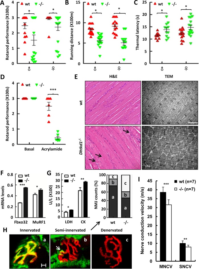
Dhtkd1−/− mice develop CMT2-like phenotypes. (A to C) Tests of general motor and sensory performance using an accelerating rotarod (A), a treadmill (B), and a hot plate test (C) (n = 15 mice per genotype). (D) When mice were exposed to 400 ppm of acrylamide for 6 days, retention time was assayed using an accelerating rotarod test (n = 7 mice per genotype). (E) H&E staining (left) denotes atrophies (arrows) in Dhtkd1−/− gastrocnemius muscle. Magnification, ×200. Electron microscopy (right) of the gastrocnemius sections in wt and Dhtkd1−/− mice shows sarcomere disorder in Dhtkd1−/− gastrocnemius muscle. Scale bar, 2 μm. (F) mRNA levels of atrophy-associated ubiquitin ligases, Fbxo32 and MuRF1, detected by real-time PCR. (G) Circulating LDH and CK levels in serum (n = 6 mice per genotype). (H) Colocalization of alpha-bungarotoxin and SV2/SH3 markers indicates muscle innervations, partial colocalization representing semi-innervated junctions (arrow), and denervation as shown by alpha-bungarotoxin staining. On the basis of this classification, the percentages of innervated, semi-innervated, and denervated NMJ were analyzed by a chi-square test (P < 0.001). Scale bar, 5 μm. (I) Sciatic nerve conduction velocity. Values in panels A to D are shown as means ± SEM, while values shown in panels F, G, and I are shown as means ± SD. A Student two-sided t test was used for data in panels A to D, F, G, and I. *, P < 0.05; **, P < 0.01; ***, P < 0.001.

