FIG 4.
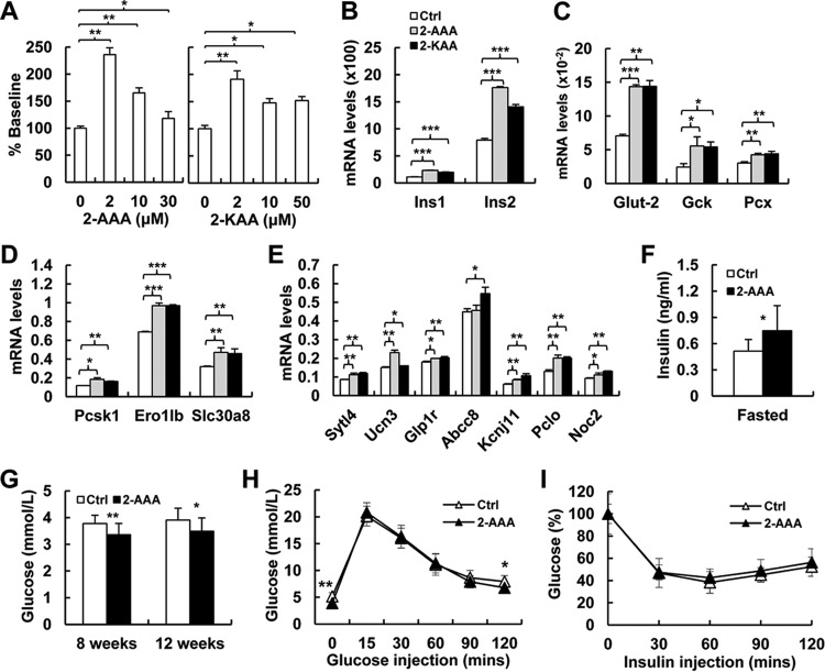
2-AAA and 2-KAA induce insulin biosynthesis and secretion in vitro and in vivo. (A) The effects of 2-AAA (19) and 2-KAA on insulin secretion in primary mouse islets from wt mice. (B) Real-time PCR analysis shows significant increases in Ins1 and Ins2 expression in Dhtkd1−/− islets treated with 2-AAA and 2-KAA. (C) Real-time PCR analysis of genes involved in glycolytic flux of islets treated with 2-AAA and 2-KAA. (D) Real-time PCR analysis of Dhtkd1−/− islets reveals increased expression of genes involved in insulin biosynthesis in 2-AAA- and 2-KAA-treated mice compared to levels in the wt. (E) Real-time PCR analysis of genes related to different insulin secretion pathways after 2-AAA and 2-KAA treatment shows elevated levels of these genes in Dhtkd1−/− islets. (F) Fasting plasma insulin was measured after 8 weeks of 2-AAA intake (2.5 mg/ml drinking water; n = 9 mice per group) (19). (G) Decreased fasting glucose levels in mice after 8 weeks and 12 weeks of 2-AAA intake are shown (n = 9 mice per group). (H) GTT was performed on 16-week-old control and 2-AAA-fed mice after 16 h of fasting using 2 g of glucose/kg body weight by i.p. injection. Plasma glucose levels during the GTT are presented. (I) ITT was performed on 6-h-fasted 16-week-old control and 2-AAA-fed mice with i.p. injection of insulin at 0.75 unit/kg body weight. Plasma glucose levels are presented as percent change from the glucose level at time zero. A Student two-sided t test was used, and values are shown as means ± SD. *, P < 0.05; **, P < 0.01; ***, P < 0.001.

