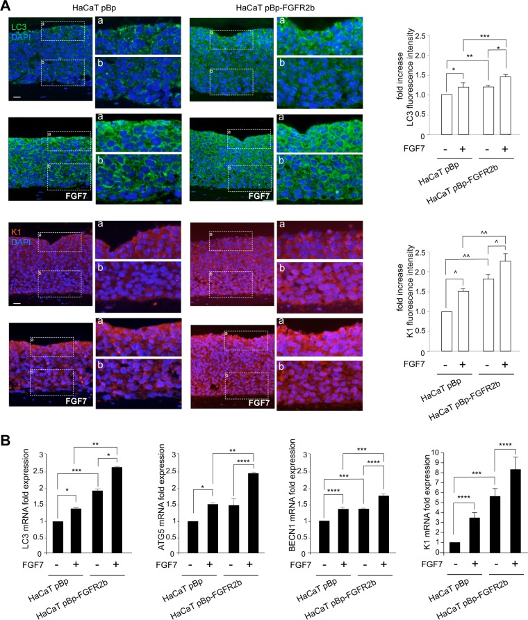
FIG 1.
FGFR2b expression and signaling trigger both autophagy and epidermal early differentiation. Organotypic skin equivalents using HaCaT pBp and pBp-FGFR2b clones, prepared as reported in Materials and Methods, were grown in complete medium and then left untreated or stimulated with FGF7 for an additional 4 days. (A) Quantitative immunofluorescence analysis shows that, in pBp rafts stimulated with FGF7, LC3 and K1 staining appear intense starting from the first suprabasal layer. In HaCaT pBp-FGFR2b rafts, both stains are already detectable in the basal layer and are increased compared to the levels in controls, particularly after FGF7 stimulation. Quantitative analysis of the fluorescence intensities was performed as described in Materials and Methods, and the results are expressed as fold increases with respect to mean values ± standard errors (SE) for pBp cells. Student's t test was performed, and significance levels are defined as P values of <0.05. For comparison to the results for the corresponding FGF7-unstimulated cells: *, P < 0.01; ^, P < 0.001. For comparison to the results for the corresponding pBp cells: **, P < 0.0001; ***, P < 0.05; ^^, P < 0.001. Bar = 25 μm. (B) Real-time RT-PCR analysis shows that mRNA expression levels of LC3, ATG5, and BECN1, as well as of K1, are significantly increased upon FGF7 stimulation, particularly in pBp-FGFR2b rafts. Results are expressed as mean values ± SE. Student's t test was performed, and significance levels are defined as P values of <0.05. For comparison to the results for the corresponding FGF7-unstimulated cells: *, P < 0.01; ****, P < 0.05. For comparison to the results for the corresponding pBp cells: **, P < 0.01; ***, P < 0.05.

