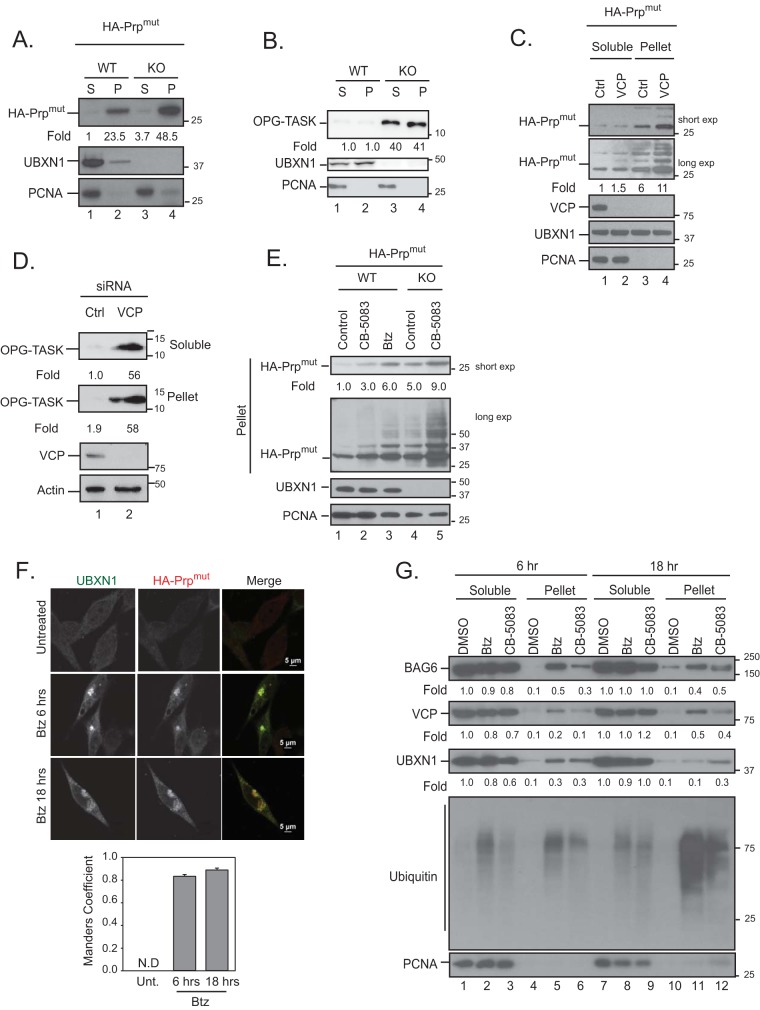
FIG 7.
Loss of VCP-UBXN1 leads to the accumulation of HA-PrPmut in insoluble compartments. (A and B) Wild-type or UBXN1 KO cells were transiently transfected with HA-PrPmut (A) or OPG-TASK (B) and fractionated into soluble (S) and insoluble pellet (P) fractions. Levels of the substrate in each compartment were probed. (C and D) HeLa Flp-in T-REX cells were transfected with control or VCP siRNA and HA-PrPmut (C) or OPG-TASK (D) and fractionated as described above for panel A. (E) Wild-type and UBXN1 KO cells were transfected with HA-PrPmut and treated with 5 μM CB-5083 or 1 μM bortezomib (Btz) for 4 h. The levels of PrP were determined. (F) HA-PrPmut was transfected into HeLa Flp-in T-REX cells, and cells were treated with 1 μM bortezomib for 6 or 18 h. Cells were fixed, stained for endogenous UBXN1 and HA, and imaged. Colocalization was determined by the Manders overlap coefficient for 25 cells in 3 replicate experiments. N.D, not determined. (G) HeLa Flp-in T-REX cells were treated with 5 μM CB-5083 (VCP inhibitor) or 1 μM bortezomib for 6 or 18 h. Cells were fractionated into soluble and pellet fractions, and the levels of VCP, UBXN1, and BAG6 in each fraction were determined. Fold changes for all panels were determined by densitometry and normalized to PCNA levels (n = 3).

