FIG 9.
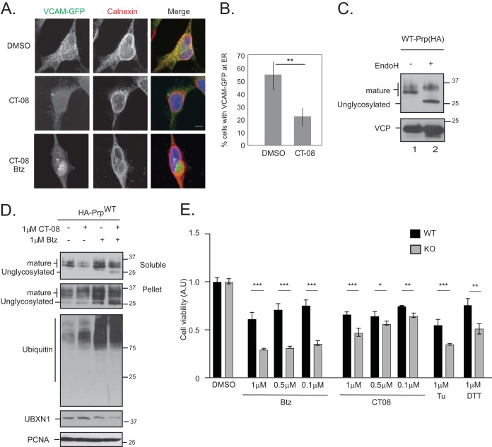
Loss of UBXN1 sensitizes cells to proteotoxic stress. (A) HeLa Flp-in T-REX cells were transfected with VCAM1-GFP and treated with 100 nM CT08 for 24 h. A total of 1 μM bortezomib was added during the final 8 h of CT08 treatment. Cells were fixed, stained for calnexin, and imaged. In CT08-treated cells, VCAM-GFP no longer localized to the ER. Cotreatment with bortezomib and CT08 was cytotoxic, and few cells remained to be quantified; however, in surviving cells, VCAM-GFP was localized to an aggresome-like structure next to the nucleus. Data for 50 cells were quantified in each of 2 independent experiments. (B) Quantification of data in panel A. (C) To determine the migration of mature and unglycosylated wild-type PrP, cell lysates expressing HA-PrP were treated with EndoH and resolved on SDS-PAGE gels. (D) Wild-type HA-PrP-expressing HeLa Flp-in T-REX cells were treated with 1 μM CT08 or 1 μM bortezomib for 6 h, as indicated. Cells were fractionated into soluble and pellet fractions. Mature and unglycosylated PrPs are indicated. (E) Wild-type and UBXN1 KO cells were plated in triplicate into 96-well plates and treated with the indicated concentrations of bortezomib, CT08, DTT, or tunicamycin (Tu) for 24 h. Cell viability was measured and normalized to the value for DMSO-treated controls for each cell line. **, P ≤ 0.01 (as determined by one-way ANOVA) (n = 2).

