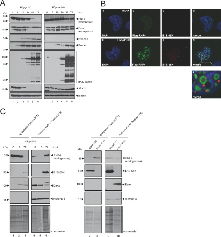
FIG 1.
HAdV-mediated modulation of RNF4 protein during infection. (A) H1299 cells were infected with either wt virus (H5pg4100, left) or with an E1B-55K null mutant (H5pm4149, right) at a multiplicity of 50 FFU per cell and harvested after indicated time points postinfection. Total cell extracts were prepared with low-salt RIPA buffer, separated by SDS-PAGE, and subjected to immunoblotting using RNF4 mouse MAb (kindly provided by Takeshi Urano), Daxx rabbit pAb 07-471 (Upstate), mouse MAb 2A6 (α-E1B-55K), E4orf6 mouse MAb RSA3, HAdV-5 rabbit polyclonal serum L133, Mre11 rabbit pAb pNB 100-142 (Novus Biologicals, Inc.), rabbit MAb α-E2A (α-E2A/DBP), and MAb AC-15 (anti-β-actin) as a loading control. Molecular sizes, in kDa, are indicated on the left, with relevant proteins on the right. (B) H1299 cells transfected with 2 μg pFlag-RNF4-WT, infected with wt virus (H5pg4100) at a multiplicity of 20 FFU per cell, and fixed with 4% PFA after 48 h postinfection. Cells were labeled with anti-Flag mouse MAb M2 (Sigma-Aldrich, Inc.), detected with Alexa 488 (α-Flag; green) and mouse MAb 2A6 (α-E1B-55K), detected with Cy3 (α-E1B-55K; red)-conjugated secondary antibody. Nuclei are labeled with DAPI (4,6-diamidino-2-phenylindole). Representative α-Flag (green; Bb, Bf), α-E1B-55K (red; Bc, Bg), and DAPI (blue; Ba, Be) staining patterns, an overlay of the single images (merge; Bd, Bh), and an enlarged overlay (merge; Bi) are shown (magnification, ×7,600). (C) H1299 cells were infected with wt virus (H5pg4100, left and right) or an E1B-55K null mutant (H5pm4149, right) at a multiplicity of 50 FFU per cell and harvested after indicated time points postinfection (left) or after 48 h (right). Cell extracts were fractionated into cytoplasm and insoluble nuclear factions. Equivalent amounts of protein for each fraction were separated by SDS-PAGE and subjected to immunoblotting using the Ab indicated in panel A plus rabbit MAb H3 (α-histone 3). Molecular sizes, in kDa, are indicated on the left, and relevant proteins are on the right.

