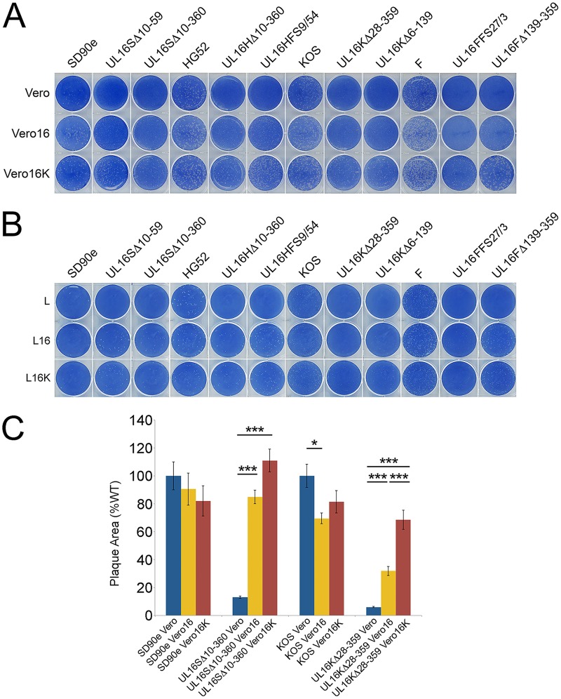
FIG 4.
Reciprocal complementation between HSV-2 and HSV-1 UL16 proteins. (A and B) Monolayers of Vero, Vero16, and Vero16K cells (A) or L, L16, and L16K cells (B) were infected with identical dilutions of each HSV-2 and HSV-1 UL16 mutant. The cells were fixed and stained with 0.5% methylene blue in 70% methanol at 72 hpi. (C) Vero, Vero16, or Vero16K cells were infected with the indicated viruses and fixed, and plaques were stained using antiserum against HSV Us3 by indirect immunofluorescence microscopy at 24 hpi. Plaque sizes were determined as described in Materials and Methods (n = 40 plaques per strain). The error bars represent standard errors of the mean. HSV wild-type strains SD90e and KOS were normalized to 100%. *, P < 0.05; ***, P < 0.0001.

