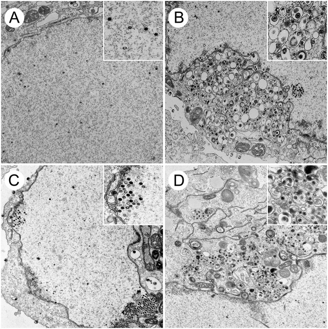
FIG 6.
Ultrastructural analysis of HSV-1-infected cells. Vero cells were infected with HSV-1 F (A and B) and the UL16 deletion mutant, UL16FΔ139-359 (C and D), at an MOI of 3. At 16 hpi, cells were fixed and processed for TEM as described in Materials and Methods. (B) Nonenveloped cytoplasmic capsids and enveloped virions can be observed in the cytoplasm of F-infected cells. (D) Enveloped virions were less frequently observed in the cytoplasm of UL16FΔ139-359-infected cells, where nonenveloped capsids were abundant. (A and C) Nuclear capsids were readily detected in the nuclei of both F-infected cells (A) and UL16FΔ139-359-infected cells (C).

