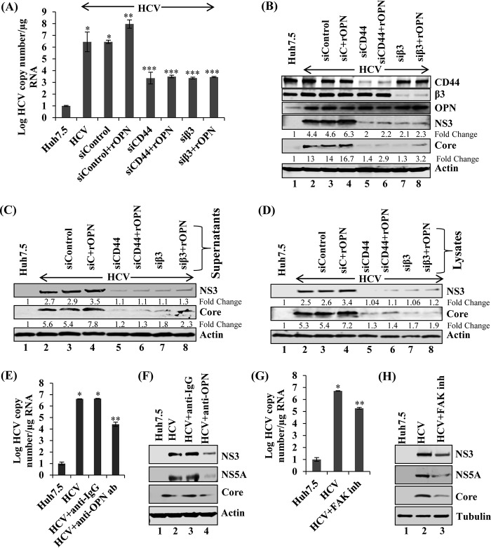
FIG 2.
Exogenous recombinant OPN (rOPN) stimulates HCV replication, infectivity, and assembly. (A) HCV-infected Huh7.5 cells (day 4) were transfected with sicontrol, siCD44, and siβ3. At 24 h post-siRNA transfection, cells were incubated with rOPN (50 nM) for another 48 h. Total RNA was extracted and HCV copy number was analyzed using quantitative RT-PCR. Data represent means ± SDs from three independent experiments performed in duplicate. *, P < 0.05 compared to mock-infected Huh7.5 cells; **, P < 0.01 compared to HCV-infected Huh7.5 cells transfected with sicontrol; ***, P < 0.001 compared to HCV-infected Huh7.5 cells transfected with sicontrol. (B) Equal amounts of cellular lysates from panel A were immunoblotted with the indicated antibodies. (C) The cell culture supernatants collected at various conditions described for panel A were incubated with naive Huh7.5 cells for 6 h, and then cells were washed and replaced with fresh medium. At day 3 postinfection, cellular lysates were immunoblotted with the indicated antibodies. (D) The cells from panel A were suspended in DMEM with 10% fetal calf serum (FCS), lysed by freeze-thaw cycles (4 times) on dry ice and a 37οC water bath, and centrifuged at 4,000 rpm for 5 min. The supernatants were incubated with naive Huh7.5 cells for 6 h and replaced with fresh media. At day 3 postinfection, equal amounts of cellular lysates were immunoblotted with the indicated antibodies. Tubulin was used as a protein loading control. (E) HCV-infected Huh7.5 cells (day 6) were incubated with anti-OPN (1:100) and control isotype goat IgG antibodies. At 24 h posttreatment, total cellular RNA was extracted and HCV copy number was analyzed by quantitative RT-PCR. The values are the means ± SDs from three independent experiments performed in duplicate. *, P < 0.05 compared to mock-infected cells (Huh7.5); **, P < 0.01 compared to HCV-infected Huh7.5 cells neutralized using control IgG antibody. (F) Equal amounts of cellular lysates from panel E were subjected to Western blot analysis using anti-NS5A, anti-NS3, and anti-core antibodies. (G) HCV-infected Huh7.5 cells (day 6) were incubated with FAK inhibitor (inh) PF573228 (2 μM). At 12 h posttreatment, total cellular RNA was extracted and HCV copy number was analyzed by quantitative RT-PCR. The values are means ± SDs from three independent experiments performed in duplicate. *, P < 0.05 compared to mock-infected cells (Huh7.5); **, P < 0.01 compared to HCV-infected Huh7.5 cells treated with an equal amount of DMSO. (H) The cellular lysates from panel G were immunoblotted with the indicated antibodies. Actin and tubulin were used as protein loading controls.

