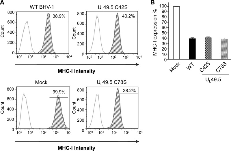
FIG 10.
FACS analysis of MHC-I cell surface expression in wt and UL49.5 mutant virus-infected MDBK cells. Cells were infected with wt, C42S, and UL49.5 C78S mutant viruses. MHC-I cell surface expression was detected at 18 hpi with a monoclonal anti-MHC-I antibody in combination with an anti-mouse FITC antibody and determined by FACS analysis. (A) Representative graphs of MHC-I cell surface expression of virus-infected cells. (B) Mean of three independent experiments of MHC-I expression in virus-infected cells.

