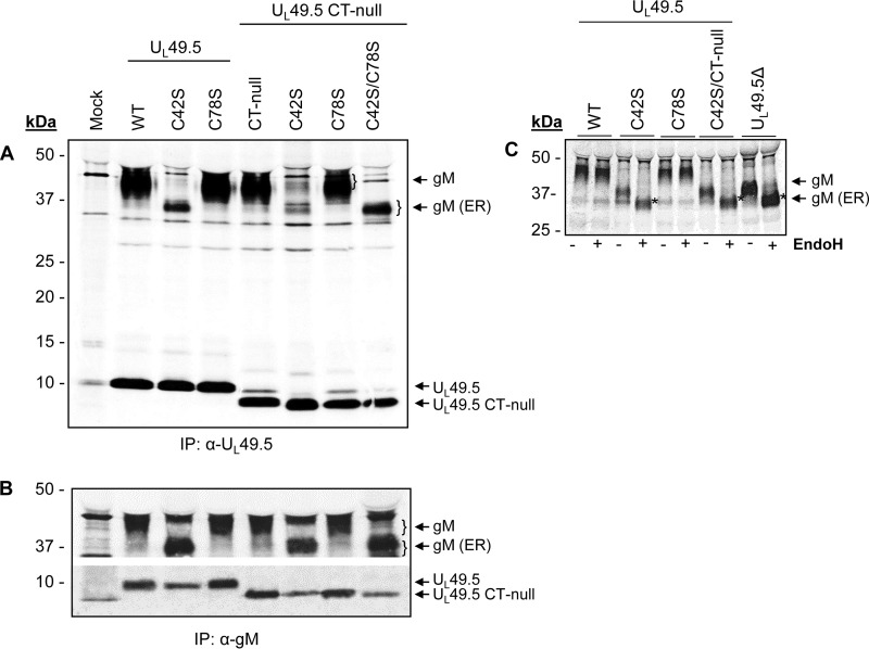
FIG 4.
Analysis of UL49.5-gM interaction by radioimmunoprecipitation assay. 35S-labeled lysates from mock-infected or BHV-1 UL49.5 mutant virus-infected MDBK cells were immunoprecipitated with anti-UL49.5-specific (A) or anti-gM-specific (B) polyclonal antibodies, separated by SDS-PAGE, and visualized by autoradiography. Note that there is a nonspecific 43-kDa faint band in the mock-infected sample in both panels A and B; this band is also present in the wt- and mutant virus-infected lysate samples but is visible only when the gM (43 kDa) is not processed (C42S mutants). Also, in panel A anti-UL49.5 antibody precipitated a nonspecific 9-kDa faint band in the mock-infected sample, and this band is also visible in the CT-null lysates. (C) 35S-labeled lysates from various mutant virus-infected MDBK cells were immunoprecipitated with anti-gM-specific antibody and digested with EndoH (+). The untreated samples (−) were included as controls. EndoH-sensitive, immature gM is marked by asterisks.

