FIG 1.

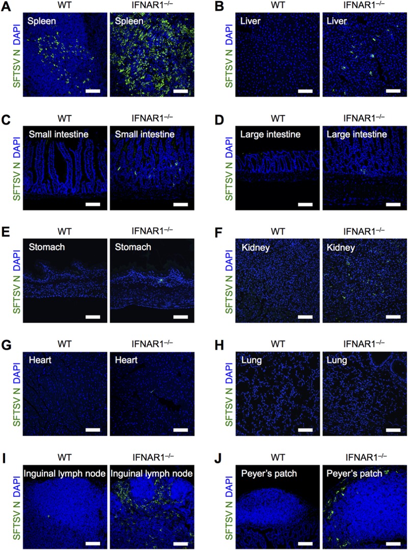
Deficiency of IFN-I signaling promotes SFTSV replication in secondary lymphoid tissues. Shown is immunohistochemical analysis of SFTSV N protein in tissues from WT and IFNAR1−/− mice at 2 dpi. Spleen (A), liver (B), small intestine (C), large intestine (D), stomach (E), kidney (F), heart (G), lung (H), inguinal lymph node (I), and Peyer's patch (J) sections were costained for SFTSV N (green) and DAPI (blue). All the sections were analyzed by confocal microscopy. Scale bars, 100 μm. Representative images from 3 mice are shown.
