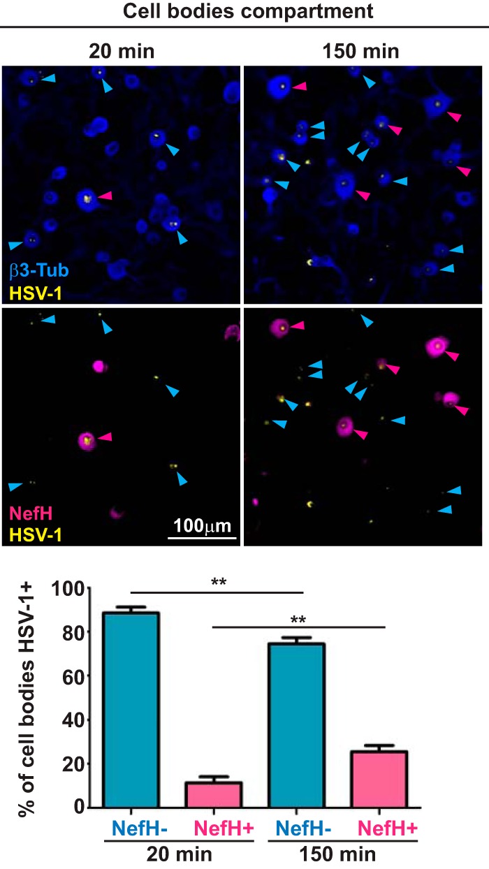
FIG 3.

HSV-1 early-infection efficiency is lower in NefH+ neurons. TG neurons cultured in microfluidic devices for 3 days were infected synchronously at the distal axon compartment with KOS (MOI = 200). Cultures were fixed and processed at two time points: 20 min (left) and 150 min (right) postadsorption. Immunostaining for β3-Tub (blue) and NefH (magenta) revealed total neurons and NefH+ neurons, respectively. Immunostaining for HSV-1 is in yellow. Blue arrowheads point to NefH−-infected neurons. Magenta arrowheads point to NefH+-infected neurons. The graph represents the percentage of NefH− and NefH+ cell bodies positive for KOS at the early and late time points. The threshold for KOS detection was set using mock-infected chambers. Data are for 7 chambers for 20 min postadsorption and 7 chambers for 150 min postadsorption from 2 independent experiments. **, P < 0.01.
