
FIG 6.
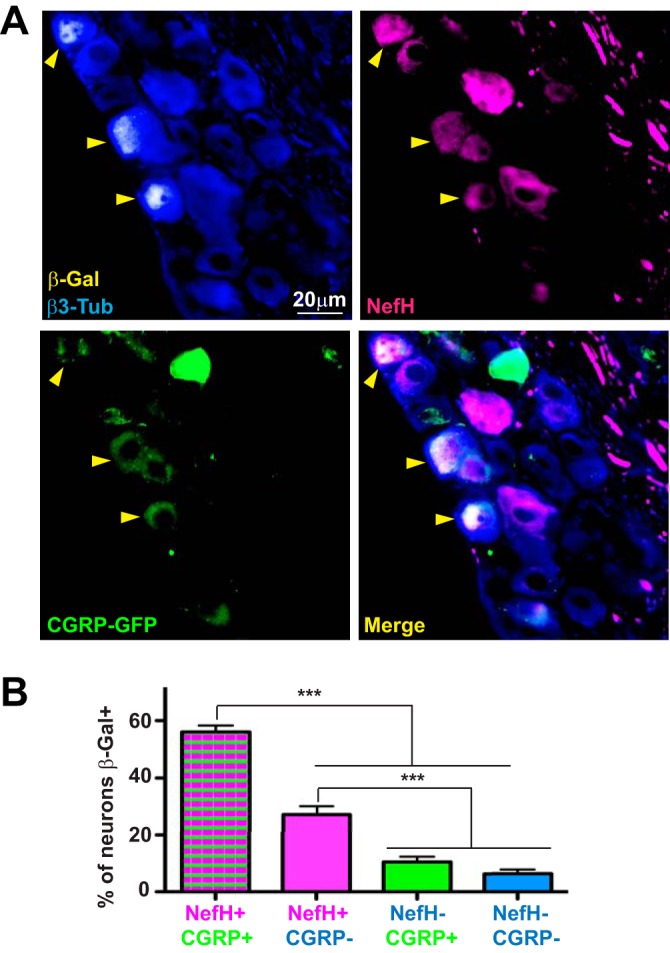
HSV-1 LAT expression is predominantly observed in NefH+ CGRP+ sensory neurons following corneal infection. (A) Representative image of TGs from adult CGRP-GFP mice corneally infected with KOS/62 (1 × 10⁶ PFU/eye) for >21 days. Tissue sections were immunostained for β-Gal (yellow), β3-Tub (blue), and NefH (magenta). CGRP+ neurons were green owing to GFP expression from the transgenic mouse. Yellow arrowheads point to β-Gal+ neurons in which LAT promoter activity is evident. The merged image is shown at the bottom right. (B) The graph represents the percentage of each subpopulation of neurons positive for β-Gal. The threshold for β-Gal detection was set using mock-infected tissue. No fewer than 6 random sections per TG were analyzed. Data are for 12 TGs from 2 independent experiments. ***, P < 0.001. Bar, 20 μm.