
Fig. 2. The decussating and nondecussating DRTTs.
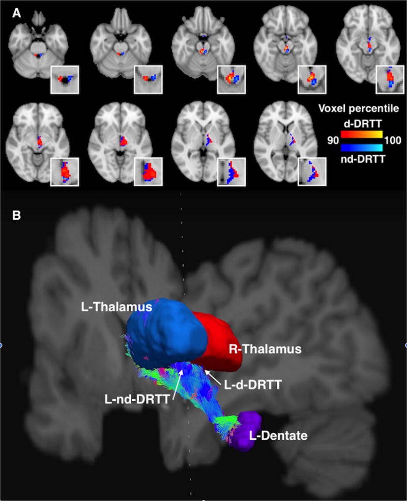
A. Axial slices showing the d-DRTT (red) and nd-DRTT (blue), thresholded to include the top 10% of nonzero voxels by structural connectivity for each tract. For clarity, only the DRTT tracks arriving at the left thalamus (i.e. right decussating and left nondecussating tracts) are depicted. Figure has been up-sampled to 0.5mm isotropic resolution for better visualization clarity. Insets are enlargements of the same axial slices. B. A 3-dimensional visualization of the DRTT pathways originating in the left dentate nucleus and arriving at the ipsilateral (blue) and contralateral (red) thalamus. View is from the posterior aspect, with coronal and saggital slices of T1-weighted image shown for reference. Streamline coloration is based on fiber orientation, where blue is superior-inferior, green is anterior-posterior, and red is left-right, per convention.