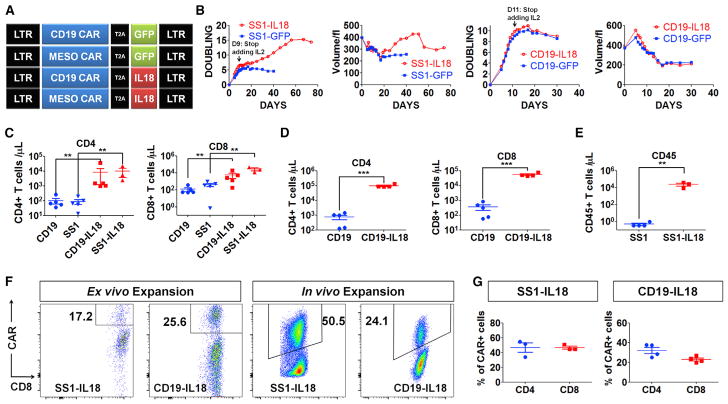
Figure 1. IL18-CAR T Cells Have Enhanced Proliferation In Vitro and In Vivo.
(A) The construct designs.
(B) Population doubling and cell volume of SS1 anti-mesothelin or anti-CD19 CAR T cells following the first restimulation with irradiated K562-Meso or K562-CD19 with exogenous IL-2.
(C) NSG mice (n = 5) bearing an AsPC1 pancreatic flank tumor received CD19 or SS1 CAR T cells (2e6), and 3 weeks later, peripheral blood was analyzed by Trucount.
(D) NSG mice (n = 5) bearing a systemic Nalm6 acute lymphoblastic leukemia (ALL) tumor received 1e6 CD19 or CD19-IL-18 CAR T cells. After 18 days, circulating T cells were assessed.
(E) Tumor-free NSG mice (n = 5) were inoculated with 5e6 SS1 or SS1-IL-18 CAR T cells and, 3 weeks later, analyzed for circulating T cells.
(F and G) CAR T cells were analyzed on day 9 of ex vivo expansion. In vivo expansion of SS1-IL-18 and CD19-IL-18 CAR T cells was determined by harvesting spleens from the mice described in (C) and (D), respectively.
(F) Representative fluorescence-activated cell sorting (FACS) plots of CD8+CAR+ cells.
(G) The percentage of CD4+CAR+ or CD8+CAR+ T cells in spleens from (F).
All data with error bars are presented as mean ± SEM. Student’s t test: **p < 0.01, ***p < 0.001.

