
Fig. 1. Circulating tumor cells (CTCs) that transited through the lymph node detected in mouse models.
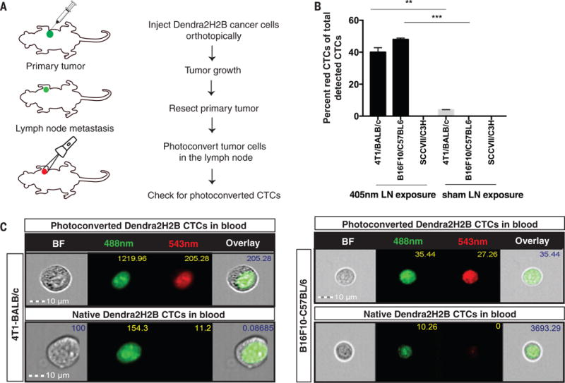
(A) Dendra2H2B positive cancer cells were injected orthotopically into syngeneic recipients. Approximately 20 days later, the primary tumor was resected and tumor draining lymph nodes were photoconverted using a 405nm diode for 5 consecutive days. Blood was analyzed for the presence of green/red fluorescent CTCs using an Amnis Imagestream flow cytometer. (Photoconverted animals: 4T1 model n=11, B16F10 model n=7, SCCVII model n=5. Control animals: 4T1 model n=5, B16F10 model n=7, SCCVII n=3) (B) Dendra2H2B-4T1 cells and Dendra2H2B-B16F10 cells but not Dendra2H2B-SCCVII cells photoconverted in the draining lymph node were detected in the blood. Data are represented as the percentage of red CTCs (photoconverted) to total detected CTCs. Green CTCs were detected in all three models. *p<0.05 comparing 405nm light exposure to sham exposure for individual cells lines. (C) Representative images obtained by Imagestream of CTCs from 4T1/BALB/c and B16F10/C57BL/6 mouse models show positive photoconverted cancer cells verified by nuclear localization of Dendra2H2B. Scale bar=10μm.