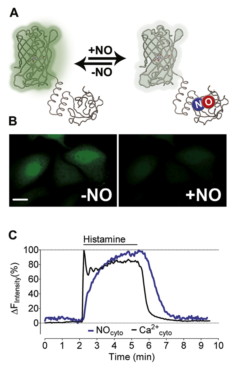
Fig. 1. Simultaneous imaging of Ca2+ and NO in single endothelial cells.
(A) The scheme represents the functional principle of G-geNOp that FP fluorescence is quenched upon NO binding to the probe. (B) Representative wide field images of EA.hy926 cells expressing G-geNOp prior to exposure (left panel) and in the presence of the NO-donor NOC-7 (10 μM, right panel). (C) Representative [Ca2+]cyto (black curve) and [NO]cyto (blue curve) signals shown in percentage of the respective maximal increases in response to 100 μM histamine. Curves represent at least 4 independent experiments. (For interpretation of the references to colour in this figure legend, the reader is referred to the web version of this article.)

