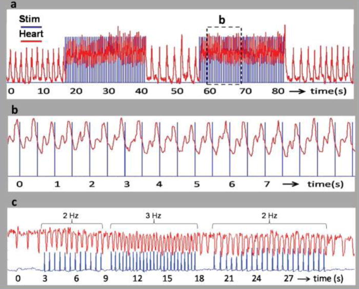
Figure 2. Pacing of the embryonic quail heart from Jenkins et al16.

a,b, The trigger pulse (blue) from the pulsed laser is superimposed on the heart rate (red). (a) Recording from a stage 17 (59 hour) quail embryo paced at 2 Hz. The laser pulse duration was 1 ms and the radiant exposure was 0.92 J/cm2 per pulse. The laser pulses were turned on and off several times to demonstrate the robustness of optical pacing. The heart rate increased from 0.634 to 2 Hz when the stimulation laser pulses were started and decreased to 0.693 Hz by the end of the trace in 2a. (b) The dashed box in 2a is expanded for a close up view in 2b. Clearly the trigger pulse and LDV signal were synchronized with each laser pulse eliciting a heartbeat. (c) Recording from a stage 14 (53 hour) quail embryo. The laser pulse duration was 2 ms and the radiant exposure was 0.84 mJ/cm2 per pulse. The frequency of the laser pulses were varied from 2 Hz to 3 Hz and back to 2 Hz to demonstrate the ability of the embryo heart to follow the pulse frequency.
