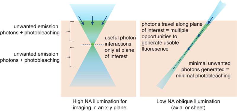
Figure 2.
Comparing high-NA point-scanning with low-NA light sheet or axial beam illumination. High NA confocal microscopy illuminates tissue above and below the focal plane causing photodamage to regions that are not being imaged, while requiring a pinhole to reject light originating from these regions. Axial illumination restricts light to only the plane / line being imaged, reducing both background and photodamage to regions that are not being imaged. However, a further benefit of axial beams is that the beam or sheet is composed of photons traveling in their direction of propagation. As a result, photons forming the sheet have repeated opportunities to interact with fluorophores along their path, whereas high NA confocal imaging only detects interactions occurring at the focal plane. This effect makes axially-extended beams more efficient – generating more usable fluorescent photons per photon entering the tissue.

