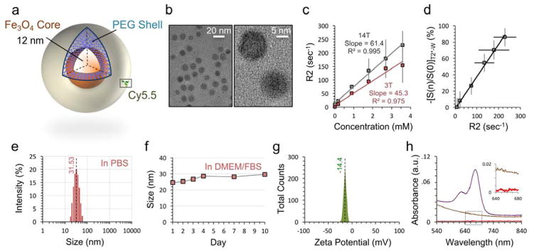
Figure 1. Characterization of iron oxide nanoparticles.
a, Schematic of nanoparticle with metallic 12 nm iron oxide core and amine-terminated PEG shell. The core and shell are joined by a cross-linked siloxane intermediate layer. Cy5.5 fluorophore is conjugated on a subset of the nanoparticles. b, TEM of the nanoparticle at low magnification (left) demonstrates monodispersity and spherical core shape. High magnification (right) displays linear features of the iron oxide crystal lattice. c, Magnetic T2 relaxivity of IOSPM NPs in PBS at 25 °C is calculated to be 45.3 s−1mM−1 at 3 T (red), and 61.4 s−1mM−1 at 14 T (grey). d, Quantitative IOSPM NP data at 14 T. NP concentrations and R2 values in (c) are plotted against respective T2*-W signal changes from baseline, demonstrating a linear relationship between these two measurement techniques. e, Dynamic light scattering (DLS) data demonstrates an average NP hydrodynamic size of 31.53 nm in PBS, with good size uniformity. f, Monitoring of NP size in DMEM/FBS reveals <5 nm change over 10 days, with a stable hydrodynamic size of ~29 nm reached between days 4–10. g, Zeta-potential measurement shows a net-negative particle surface charge of −14.4 mV. h, Confirmatory biodistribution experiments in mice required fluorophore labeling with Cy5.5. UV-visible spectra for NP samples prior to Cy5.5 conjugation (brown) and after conjugation and purification (purple) confirm presence of the fluorophore (UV-vis baseline was PBS, and samples of equivalent Fe3O4 concentration are shown). The aqueous buffer from the purified Cy5.5-labeled sample was extracted by centrifugation (30 K MW cutoff spin column), to demonstrate good binding, from an absence of Cy5.5 in the supernatant (red, inset).

