Figure 1.

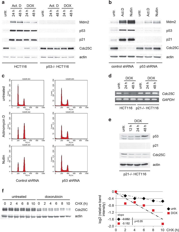
p53 regulates Cdc25C protein stability upon activation. (a) p53 wild-type and p53-null HCT116 cells were treated with 5 nM actinomycin D or 100 ng/ml doxorubicin. Twenty-four and 48 h after treatment, cell lysates were immunoblotted. (b) Stable U2OS clones expressing control or p53 shRNA were treated with 5 nM actinomycin D or 2.5 μM nutlin-3. Forty-eight hours after treatment, cell lysates were immunoblotted. (c) Cells treated as in B were stained and cell cycle profile was analyzed by flow cytometry. (d) Wild-type and p21-null HCT116 cells were treated with 500 ng/ml doxorubicin and subjected to RT–PCR analysis at the indicated time points. (e) p21-null HCT116 cells were treated as in (d). Twelve, 24 and 48 hours after treatment, cell lysates were immunoblotted. (f) HCT116 cells were treated with 100 ng/ml doxorubicin when indicated. Twenty-four hours after treatment, cells were treated with 100 μg/ml cycloheximide and harvested at the indicated time points. Normalized Cdc25C levels quantified by densitometry are shown. Statistical analysis of the slopes using covariance analysis was performed using StatsToDo software (http://www.statstodo.com).
