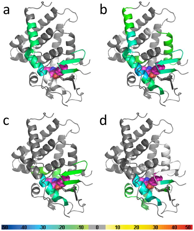
Figure 5.
Results of hydrogen-deuterium exchange mass spectrometry (HDX-MS) studies of the PXR LBD in the presence of SJB7 and SPA70. The crystal structure of the PXR LBD (PDB 5X0R) was used as the template for overlays of the differential HDX data for (a) SJB7, (b) SPA70, (c) SJB7 with the subsequent addition of SRC-1 peptide, and (d) SPA70 with the subsequent addition of NCoR peptide. The coloring of the PXR structures is based on the color bar at the bottom of the figure. Regions that were not covered are represented in white. SJB7 and SPA70 are represented as spheres, with carbon atoms depicted in pink. The SJB7 binding location is taken from the crystal structure, whereas that for SPA70 was determined by docking studies.

