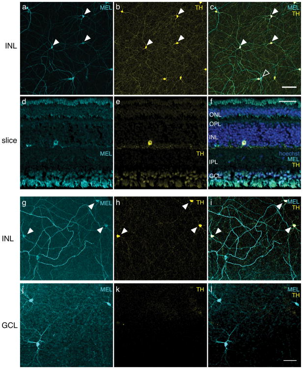
Figure 4.
Most cells immunoreactive to melanopsin (MEL) in the INL also exhibit immunoreactivity for TH. (a) MEL immunoreactivity in the INL. (b) TH immunoreactivity in the INL, same field of view as in (a). (c) Merge of (a) and (b). Eight of ten cells in the field of view were immunoreactive to anti-MEL and anti-TH; two of ten cells were immunoreactive to anti-MEL only. Closed arrowheads point to a subset of cells that are MEL and TH positive. Open arrowhead points to a cell that is only MEL+. No cells were TH+ and not MEL+. Scale bar: 100 μm. (d) Anti-MEL immunoreactivity in a transverse section. (e) Anti-TH immunoreactivity, field of view same as (d). (f) Merge of (d) and (e) with hoechst labeling. ONL, outer nuclear layer; OPL, outer plexiform layer; INL, inner nuclear layer; IPL, inner plexiform layer; GCL, ganglion cell layer. Scale bar: 50 μm. (g) MEL immunoreactivity in INL of a different retina. (h) TH immunoreactivity in the INL, same field of view as (g), but with a different TH antibody than that used in (a–f). I. Merge of (g) and (h). (j) Same field of view as (g), but focused on the GCL, showing MEL immunoreactivity. (k) Same field of view and focal plane as (j) showing TH immunoreactivity. (l) Merge of (j) and (k): Scale bar, 50 μm.

