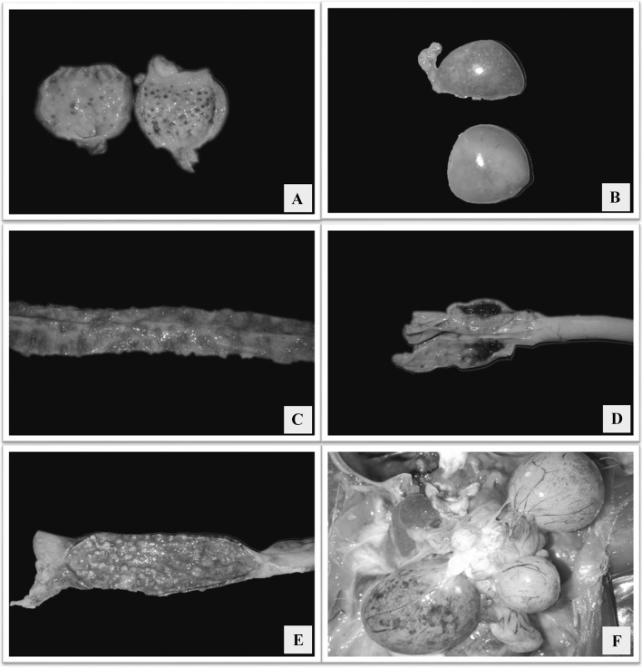
Fig. 1.
a Proventriculus showing hemorrhages in the glandular tips; b Spleen with numerous multifocal pale areas giving a mottling appearance; c Mucosa of duodenum showing multifocal to coalescing raised hemorrhagic to necrotizing ulcerated areas; d Caecal tonsils with hemorrhages; e Mucosa of rectum showing multifocal to coalescing raised necrotic ulcerated areas; f Congested ovarian follicles with petechial to ecchymotic hemorrhages

