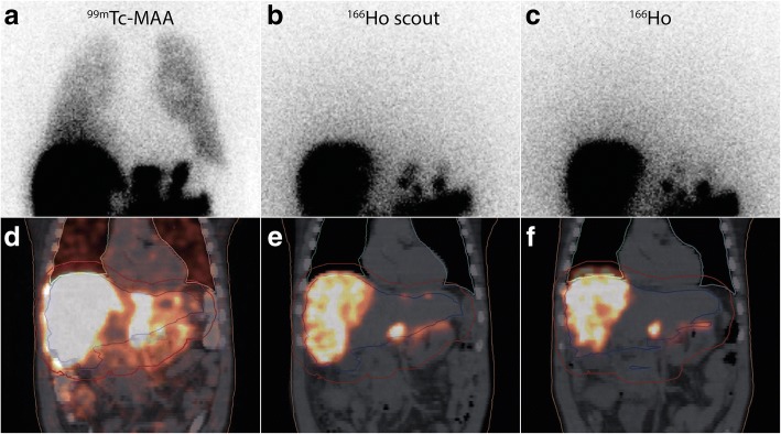
Fig. 4.
Example of lung shunt fraction overestimation by 99mTc-MAA. A patient with multiple liver metastases of a cholangiocarcinoma treated in the prior HEPAR 2 trial. Note the (visual) significant overestimation of 99mTc-MAA on planar imaging compared to the 166Ho-scout dose and 166Ho-treatment dose. Quantification of the lung shunt fraction on planar and SPECT/CT imaging confirmed the visual assessment: a 99mTc-planar = 13.4%, d 99mTc-SPECT = 6%, all 166Ho imaging modalities (b, c, e and f) with the scout dose and with the treatment dose showed a lung shunt fraction of < 1%

