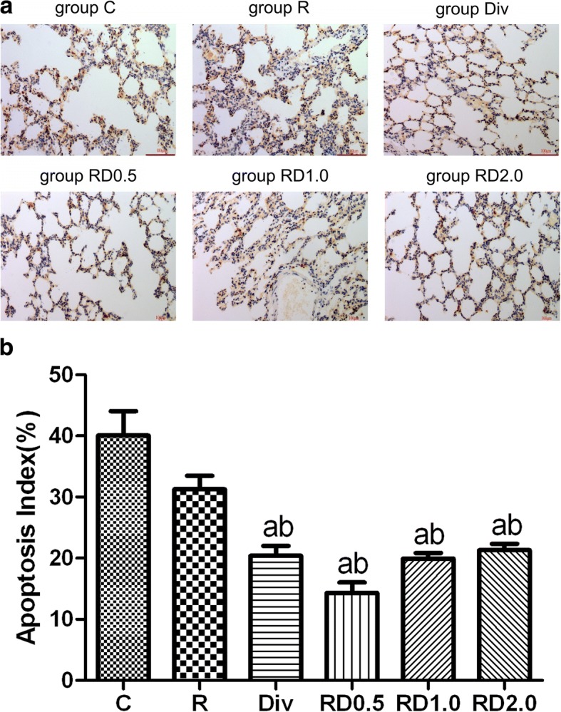
Fig. 4.

Representative images from the TUNEL apoptosis assay following one-lung ventilation. Images from the TUNEL apoptosis assay following one-lung ventilation (Fig. 4a. Yellow-brown staining indicates apoptosis. As shown in Fig. 4b the AI index was the highest in C and R groups. The AI index was significantly lower in the Div, RD0.5, RD1.0 and RD2.0 groups than in the C and R groups. Magnification × 400. C, normal saline; R, ropivacaine; Div, intravenous DEX; RD0.5, 0.5 μg/kg DEX as an adjuvant to ropivacaine TPVB; RD1.0, 1.0 μg/kg DEX as an adjuvant to ropivacaine TPVB; RD2.0, 2.0 μg/kg DEX as an adjuvant to ropivacaine TPVB
