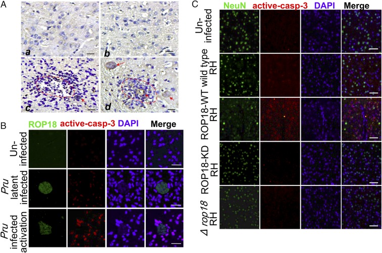
Fig. 1.
ROP18 kinase activity is required for neural apoptosis. (A) Immunohistochemical staining of ROP18 in brain sections of mice infected with the cyst-forming Pru strain reactivated by cyclophosphamide administration. (a and b) Normal brains of the uninfected group are also included. (c and d) Necrosis and inflammatory infiltration are noted in foci of recrudescence (red dotted line), and cysts in TE (red arrows) are evident. (Scale bars, 50 μm.) (B) Immunofluorescence analysis of the brain from mice infected with the cyst-forming T. gondii Pru strain reactivated by cyclophosphamide administration. The cerebral cortex region was double-stained with ROP18 (green) and active caspase-3 (red). DAPI (blue) was used to stain the nuclei. (Scale bars, 20 μm.) (C) Immunofluorescence analysis of the brain from mice infected with ROP18-WT RH, ROP18-KD RH, ∆rop18 RH, and wild-type RH tachyzoites. The cerebral cortex S1 region was double-stained with NeuN (green, a specific marker of neuron cells) and active caspase-3 (red). DAPI (blue) was used to stain the nuclei. (Scale bars, 50 μm.)

