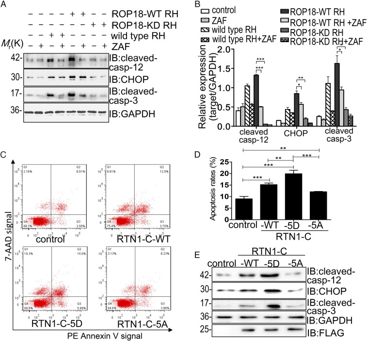
Fig. 4.
ROP18 phosphorylation of RTN1-C induces ER stress-associated apoptosis (A) After Neuro2a cells were treated with or without Z-ATAD-FMK (ZAF) for 6 h, the cells were infected with ROP18-WT RH or ROP18-KD RH tachyzoites at a multiplicity of infection of ∼3 for 24 h. Then, cell lysates were assessed by Western blotting using the indicated antibodies. GAPDH was included as a loading control. IB, immunoblot. (B) Quantitative data of A. The presented figures are from a representative study, and the quantitative data are expressed as the mean ± SD based on different assays (n = 3). *P < 0.05; **P < 0.01; ***P < 0.001 for ROP18-WT RH vs. ROP18-KD RH, ROP18-WT RH + ZAF, or RH group. (C) Neuro2a cells were transfected with FLAG-tagged RTN1-C-WT, RTN1-C-5A, or RTN1-C-5D for 24 h. Then, apoptotic cells were detected by flow cytometry after annexin V-phycoerythrin (PE)/7-aminoactinomycin D (7-AAD) staining. The plots were obtained from a representative measurement. (D) Quantitative data of C are expressed as the mean ± SD based on three different assays (n = 3). **P < 0.01 vs. negative controls; ***P < 0.001 vs. negative controls. (E) Representative Western blot analysis of cleaved caspase-12, CHOP, and cleaved caspase-3 in Neuro2a cells after transfection with FLAG-tagged wild-type RTN1-C or its mutants (RTN1-C-5D and RTN1-C-5A).

