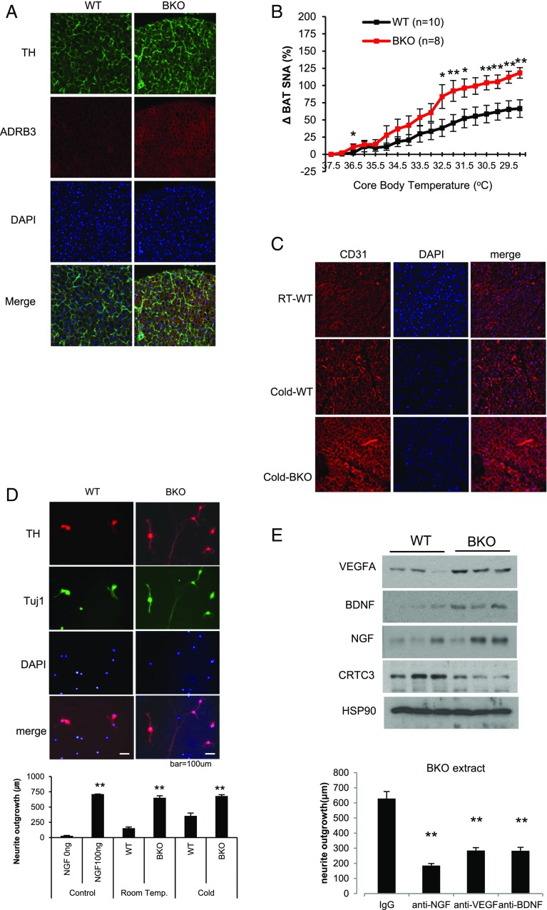
Fig. 2.
Increased sympathetic innervation and vascularization of BAT in BKO mice. (A) Immunofluorescence analysis of tyrosine hydroxylase (TH) and β3 adrenergic receptor (ADRB3) expression in BAT from WT (CRTC3 fl/fl) and BKO littermates at ambient temperature. Representative images from three independent experiments shown. (B) Sympathetic nerve activity (SNA) to BAT, measured by direct recording of nerve fibers in response to decreasing core body temperature of NCD-fed WT and CRTC3 BKO mice. n = 10 and 8 per group. Data represent means ± SEM. (C) Immunofluorescence analysis showing effect of cold exposure (15 h) on BAT vascularization in WT and BKO mice using the angiogenic marker CD31. Images are representative of three different experiments. (D, Top) Immunofluorescence analysis of cultured neurons from SCGs showing relative effects of adding WT or BKO BAT extract on neurite extension. Cells were exposed to crude extracts for 18 h and then stained with anti-TH and Tuj1 antibody to visualize sympathetic neurons. Representative images are shown. (D, Bottom) Bar graph showing relative neurite outgrowth in cells exposed to BAT extract from BKO and WT mice. Exposure to cold (3 h) indicated. n = 30–120. (E, Top) Immunoblot showing relative protein amounts for neurotrophic factors (NGF, BDNF, VEGFA) in brown-fat extracts from WT and BKO mice. n = 3 per group. (E, Bottom) Effect of anti-VEGFA, anti-BDNF, and anti-NGF neutralizing antibodies on neurite extension in response to addition of BKO BAT extract. n = 95–122 (*P < 0.05; **P < 0.01).

