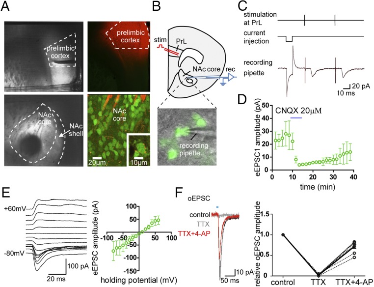
Fig. 4.
The PrL sends glutamatergic projections to D2-MSNs in the NAc core subregion. (A, Left) The PrL and the NAc could be clearly identified under the microscope in the in vitro brain-slice preparation. (Right) In brain slices prepared from Drd2-EGFP BAC transgenic mice injected with DiI into the PrL 3 wk before mice were killed, DiI-labeled fibers were found running through the NAc core and surrounding fluorescent somata representing D2-MSNs (Inset). (B) Whole-cell recordings were made from identified D2-MSNs while focal stimulation was delivered in the PrL. (C and D) A typical example showing EPSCs in response to paired stimuli (C), which could be blocked by superfusion of the AMPA receptor blocker CNQX (D); n = 4. The response to a 5-mV current injection pulse was recorded to monitor the serial resistance. (E) Current–voltage plot of eEPSCs (n = 4). (F) oEPSCs recorded in an NAc neuron with optical stimulation of ChR2-expressed prelimbic terminals in the NAc were completely abolished by TTX (1 µM) (Left) and were rescued by further 4-AP (100 µM) perfusion (Right). This phenomenon was observed consistently in eight NAc neurons. Data are presented as mean ± SEM.

