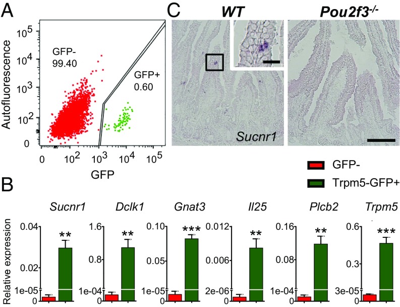
Fig. 1.
Specific expression of the Sucnr1 receptor in intestinal tuft cells. (A) Fluorescence-activated cell-sorting–based isolation of tuft cells (Trpm5-GFP–positive cells; green) and nontuft epithelial cells (GFP-negative cells; red). GFP: excitation, 488 nm; emission, 513 nm. Autofluorescence: excitation, 561 nm; emission, 585 nm. Numbers indicate percentages of cells in that population. (B) Expression of Sucnr1 and other tuft cell marker genes (Dclk1, Gnat3, Il25, Plcb2, Trpm5) in Trpm5-GFP–positive tuft cells and GFP-negative nontuft cells (control) was determined by real-time quantitative PCR. Data (mean ± SEM) are biological replicates (n = 3). **P < 0.01, ***P < 0.001 (Student’s t test). (C) In situ hybridization shows the presence of Sucnr1-expressing cells in the small intestine (jejunum) in wild-type (WT) but not in Pou2f3−/− mice. [Scale bars, 100 µm (low-magnification images) and 20 µm (Inset).]

