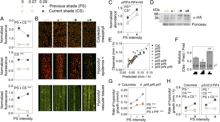
Fig. 3.
Persistent shade increases PIF4 levels to promote growth. Shown are effects of CS and PS on (A and B) nuclear fluorescence driven by the pPIF4P:PIF4-GFP transgene in mesophyll cells of the cotyledons, the epidermal and subepidermal cells of the hypocotyl (epidermis +), and the vascular tissues of the hypocotyl and on (C and D) PIF4 abundance in protein blots. (In B, scale bar = 25 μm. For representative confocal images, the GFP signal is green, while red represents autofluorescence of chlorophyll. In D, a representative protein blot is shown.) (E) Observed hypocotyl growth (SI Appendix, Table S2) vs. values predicted by a model based on PIF4 levels increased by CS and PS (i.e., overincreasing under persistent shade). (F) Goodness-of-fit of the model [as denoted by low observed (Obs)/predicted (Pred) Obs ratios and the χ2 values (numbers on bars)] when considering either stably increased, overincreasing, or decreasing PIF activity (represented in abscissa). (G) The pif4 pif5 pif7 mutant has significantly reduced response to PS. (H) Expression of PIF4 in vascular tissues promotes hypocotyl growth in Arabidopsis (both lines in the Columbia wild-type background). Means ± SE (whenever larger than the symbols) of (A and G) 20 or (H) 15 seedlings or (C) 3 biological replicates. *P < 0.05; **P < 0.01; ***P < 0.001.

