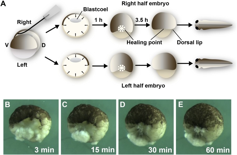
Fig. 1.
Sagittal bisection of Xenopus blastulae with an eyelash knife generates twins. (A) Diagram showing that bisection by an eyelash knife (SI Appendix, Movie S1). Bisection leaves a large wound that heals by tissue movements from all directions, closing the gap within 1 h, and leaving only a small healing point where the dorsal- and ventral-most cells become juxtaposed. Dorsal lip formation starts 3.5 h later and pigment asymmetry can be observed 1 d later in the epidermis of tailbud tadpoles. (B–E) Still images from SI Appendix, Movie S2 showing the process of convergence of blastula cell sheets toward a central healing point at which the original dorsal and ventral tissues become juxtaposed. Note that cell divisions continue in the blastula as the healing process is completed, but cell division near the wound proceeds at the same rate as in the rest of the embryo. The 1.3× objective lens of Zeiss stereomicroscope, which was connected to a DCR-PC350 Sony Handicam, was used for images.

