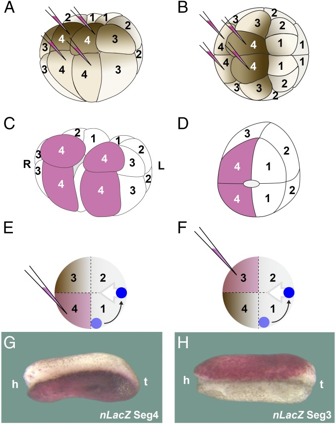
Fig. 5.
Experiment showing that the maternal pigment asymmetry in twins results from the splitting of ventral segments 3 and 4 during gastrulation; this suggests that Spemman organizer formation is displaced about 90° from its original site in the intact embryo. (A and B) Diagram indicating that segment 4 can be labeled by four injections of nLacZ mRNA at 16-cell stage in ventral and dorsal views; note that segment 3 is less pigmented than segment 4. All segments were analyzed in this way. (C) Sagittal bisection splits the embryo in two halves, allowing one to follow the fate of each segment (in the case shown here the ventral-most segment 4). (D) Diagram of a half embryo showing that during the healing process, segment 4 is juxtaposed to the dorsal-most segment 1. The healing scar is outlined in the center of half embryo. (E and F) Diagram indicating how a displacement of organizer formation (in blue) would result in the splitting of segments 3 and 4 by the involuting mesoderm (gray arrow). (G) In half embryos, the more pigmented side is derived from the segment 4 after regeneration (n = 36). (H) In bisected twins, the less pigmented epidermis is derived from segment 3 (n = 24). Note that the epidermis without lineage tracer is lighter in G than in H; both half embryos are from the same clutch and therefore the pigmentation of the epidermis is comparable. The diagrams in D–F and the embryos in G and H all correspond to right half embryos. The 40× digital magnification zoom of Axio Zoom V.16 Stereo Zoom Zeiss was used for images. h, head; L, left; R, right t, tail.

