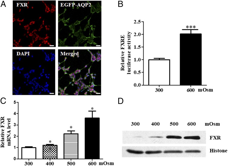
Fig. 2.
Hypertonicity-induced FXR expression in mouse MCDs. (A) Constitutive expression of FXR in mouse inner MCDs (mIMCD3). Cultured mIMCD3 cells were transfected with an EGFP-AQP2 expression vector and then exposed to hypertonic stress (600 mOsm) for 6 h. Note that FXR was mainly expressed in the nucleus (red) and AQP2 was predominantly located in cell membrane (green). (Scale bar, 25 uM.) (B) Luciferase reporter assay demonstrating that hypertonicity at 600 mOsm significantly induced FXR transcription activity. ***P < 0.001 vs. 300 mOsm (n = 12). (C) Induction of FXR mRNA expression by hypertonicity in a dose-dependent manner. Cultured mIMCD3 cells were incubated with hypertonic solutions for 6 h. *P < 0.05 vs. 300 mOsm (n = 4). (D) Western blot assay showing that hypertonicity significantly induced FXR protein expression in mIMCD3 cells. Cells were exposed to various hypertonic stress (400, 500, and 600 mOsm) for 6 h.

