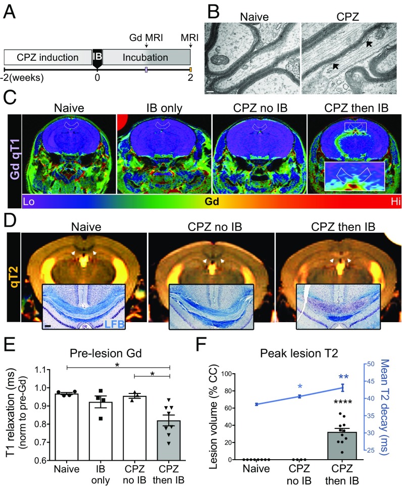
Fig. 1.
Subdemyelinating cuprizone (CPZ) triggered Gd-enhancing lesions of inflammatory demyelination. (A) Brief CPZ in an induction phase was followed by immune boost (IB) and a 2-wk incubation period on regular chow. (B) Electron micrographs of myelin ultrastructure indicate that CPZ exposure was sufficiently brief as to alter but largely preserve myelin. (C) One-week post-IB, compared with naive, IB only, or CPZ only, only the combination of abbreviated CPZ then IB resulted in significant Gd enhancement (orange/red). (D) Two-weeks post-IB, compared with controls, CPZ+IB brains alone exhibited obvious T2 white matter lesions (white arrowheads; increased signal intensity on MRI) that corresponded to inflammatory demyelination, a reaction that was termed cuprizone autoimmune encephalitis (CAE). (E) Quantification of T1 relaxation-shortening effects of brain Gd, indicative of inflammation and blood–brain barrier leakage, were observed only in CPZ+IB. (F) T2w MRI reflected the severity and extent of CAE lesions, with cases that involved nearly 60% of the anterior–posterior extent of the corpus callosum. Each data point is an individual subject and bars represent SEM. EM, n = 6; Gd MRI, n = 4 naive; n = 6 IB only; n = 3 CPZ only; n = 8 CPZ then IB. Histology, n = 12 naive controls; n = 13 IB only; n = 24 CPZ only; n = 24 CPZ then IB. Significance was determined by one-way ANOVA. *P < 0.05, **P < 0.01, ****P < 0.0001. (Scale bars: B, 100 nm; D, Insets, 100 μm.)

