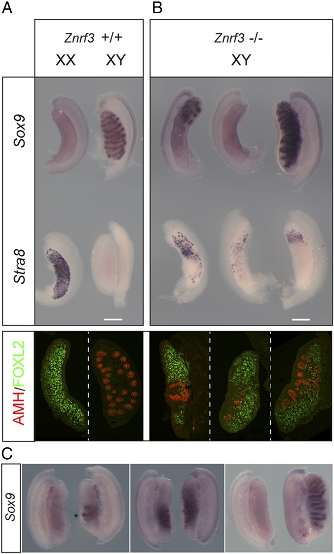
Fig. 1.
Testis determination defects in XY gonads lacking ZNRF3. (A) Wild-type controls. (B) XY gonads (14.5 dpc) lacking ZNRF3 exhibit severe morphological abnormalities in comparison with wild-type controls. WMISH with Sox9 (Top, upper row) reveals mutant gonads with a sex-reversed (ovarian) morphology (left and center) and either a major reduction in levels of Sox9 or no detectable expression. Less affected gonads have higher Sox9 levels but abnormal testis cords (right). WMISH with Stra8 (Top, lower row) reveals a significant number of meiotic germ cells in ZNRF3-deficient gonads. Gonadal sex reversal in XY gonads lacking ZNRF3 is confirmed by immunostaining of 14.5 dpc gonadal sections for AMH (red), a Sertoli cell marker, and FOXL2 (green), a granulosa cell marker (Bottom). (C) Pairs of gonads dissected from individual Znrf3 fetuses (14.5 dpc) exhibit asymmetry. Note rudimentary testis cord formation in one gonad (asterisk), but not the other (Left). Center shows distinct Sox9 expression profiles in two fetal gonads. Right shows, remarkably, an ovary on one side and a well-formed testis on the other. (Scale bar, 250 μm.)

