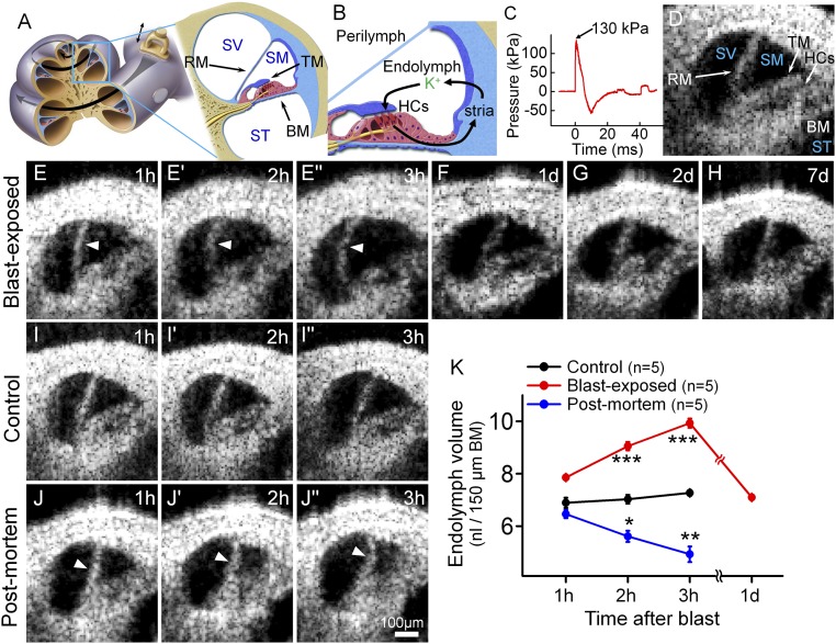
Fig. 1.
Blast exposure produces transient endolymphatic hydrops. (A) Illustration of the mouse cochlea. The apical turn we studied is expanded. BM, basilar membrane; RM, Reissner’s membrane; SM, scala media; ST, scala tympani; SV, scala vestibuli; TM, tectorial membrane. (B) Simplified schematic of K+ flow within the cochlea. K+ is secreted into endolymph by the stria vascularis (stria), taken up by transduction currents through hair cell (HCs) stereociliary bundles, and then passed back to the stria via gap junctions in supporting cells. (C) The blast wave pressure monitored by a sensor positioned just below the mouse. (D) Cross-sectional OCT image of the mouse cochlea. (E) Endolymphatic hydrops (arrowheads) progressively developed within the first 3 h after blast exposure. Images are from one representative mouse. (F–H) OCT images from different representative mice 1, 2, and 7 d after blast exposure showed normal endolymph volume. (I) OCT images from a representative live control mouse over 3 h demonstrated normal endolymph volume. (J) OCT images from an unexposed control mouse followed for 3 h after killing demonstrated progressive reductions of endolymph volume (arrowheads). (K) Scala media volume over time in blast-exposed mice, living control mice, and unexposed control mice postmortem. *P < 0.05, **P < 0.01, ***P < 0.001.

