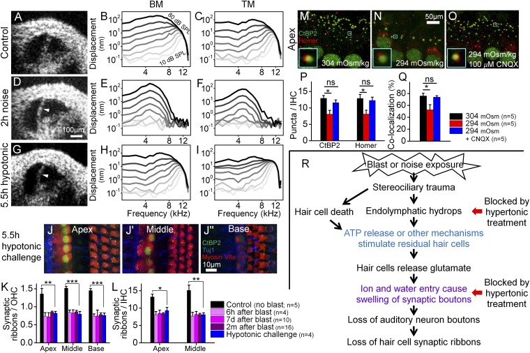
Fig. 6.
Endolymphatic hydrops correlates with cochlear synaptopathy, but not loss of hair cells or cochlear amplification. (A–C) A representative control mouse with a normal amount of endolymph had normal nonlinear vibratory responses measured from the BM and TM. (D–F) A representative mouse with endolymphatic hydrops (arrowhead) induced by 2 h of noise exposure. Vibratory responses were linear, consistent with loss of cochlear amplification. (G–I) A representative mouse with endolymphatic hydrops (arrowhead) induced by the application of hypotonic challenge to the middle ear. Vibratory responses for both the BM and TM were normal. (J) The cochlear epithelium harvested from a mouse with endolymphatic hydrops induced by hypotonic challenge to the middle ear. (K and L) Endolymphatic hydrops caused a loss of synaptic ribbons similar to that found after blast exposure. *P < 0.05, **P < 0.01, ***P < 0.001. (M–O) The cochlear epithelium harvested from representative mice perfused for 1 h with 304 mOsm (M), 294 mOsm (N), or 294 mOsm + 100 µM CNQX (O) artificial perilymph. Immunolabeling was done to visualize presynaptic ribbons in IHCs (CtBP2) and postsynaptic auditory nerve boutons (Homer). Colocalization of the pre- and postsynaptic terminals is shown (Insets). (P) Hypotonic challenge reduced CtBP2 and Homer counts. Blocking glutamate with CNQX preserved both the pre- and postsynaptic terminals. *P < 0.05; ns, not significant. (Q) Hypotonic challenge reduced the rate of CtBP2 and Homer colocalization; this effect was also inhibited with CNQX. (R) Flowchart detailing our postulated pathophysiological sequence of cochlear damage after blast exposure. The colored text reflects previously published findings [blue (4, 5), violet (6–8)]. Hypertonic challenge blocks both endolymphatic hydrops and swelling of the synaptic boutons (red arrows).

