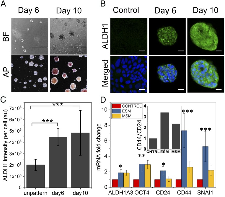
Fig. 6.
Laterally confined growth of human breast cancer cell facilitates induction of cancer stemness. (A) The bright-field images (BF) of MCF7 cells cultured on RE micropatterns up to 10 d. These MCF7 spheroids on day 10 also show alkaline phosphatase (AP) activity, unlike cells cultured on RE for 6 d. (Scale bar, 1 mm.) (B) Representative confocal micrographs of MCF7 cells grown for 6 and 10 d under lateral confinement and in control cell grown on unpatterned culture plate for 10 d. Cells were immunostained for cancer stemness marker aldehyde dehydrogenase (ALDH1) (green). (Scale bar, 20 µm.) (C) Normalized ALDH1 fluorescence intensity plot on day 6 and day 10 compared with cell grown on unpatterned culture plate for 10 d. Error bars represent SD; ***P < 0.001; Student’s t test. (D) Cancer stemness-related gene expression profile in MCF7 cells grown in RE micropatterns up to 10 d (ESM) and in MCF7 mammosphere culture (MSM) compared with cell grown on unpatterned culture plate for 10 d (CONTROL). Error bars represent SD; *P < 0.05; **P < 0.01; ***P < 0.001; Student’s t test. (Inset) The ratio of expression between CD44 and CD24.

