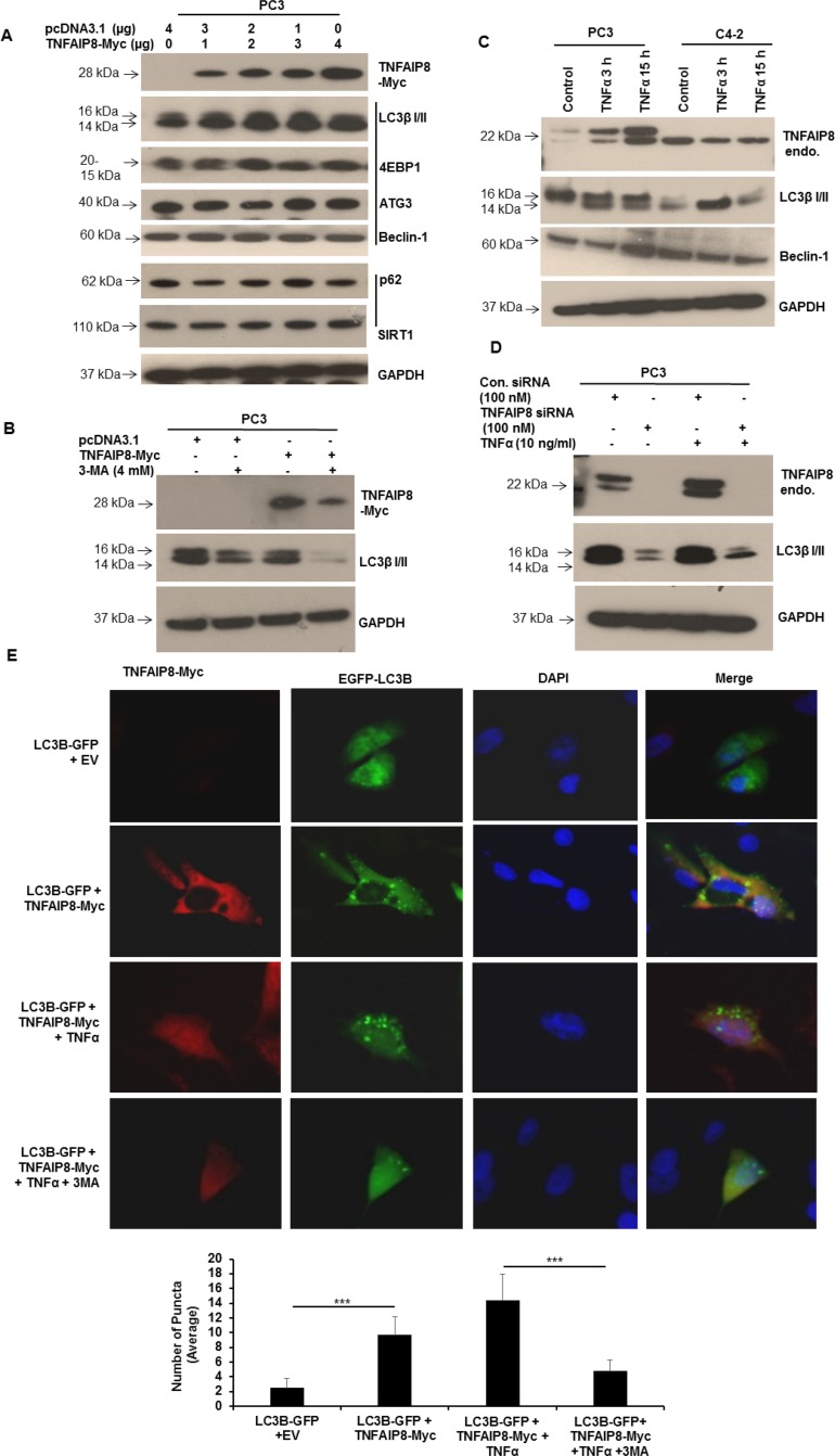
Figure 4. TNFAIP8 induces autophagy in PC3 cells.
(A) PC3 cells were transfected with empty vector or increasing amounts of TNFAIP8-Myc plasmid for 30 h. Cell extracts (50 μg) were electrophoresed using SDS-PAGE and immunoblotted with anti-Myc, anti-LC3β I/II, anti-4E-BP1, anti-ATG3, anti-Beclin 1, anti-p62, anti-SIRT1, and anti-GAPDH antibodies. (B) PC3 cells were transfected with empty vector or TNFAIP8-Myc plasmid and treated with 3-methyladenine (4 mM) for 24 h. Cell extracts (50 μg) were immunoblotted with anti-Myc, anti-LC3β I/II, and anti-GAPDH antibodies. (C) PC3 and C4-2 cells were cultured for 24 h and treated with TNFα (10 ng/ml) for 3 h and 15 h. Cell extracts (50 μg) were immunoblotted with anti-TNFAIP8, anti-LC3β I/II, anti-Beclin-1, and anti-GAPDH antibodies. Endo–endogenous. 3-MA – 3-methyladenine. (D) PC3 cells were transfected with control siRNA or TNFAIP8 siRNA (100 nM) and treated with TNFα (10 ng/ml) for 24 h as indicated. Cell extracts (50 μg) were immunoblotted with anti-TNFAIP8, anti-LC3β I/II and anti-GAPDH antibodies. Endo–endogenous. (E) PC3 cells were grown on coverslips in 6-well plates for 24 h and co-transfected with 2 μg of empty vector and GFP-tagged-LC3B plasmid or TNFAIP8-Myc plasmid and GFP-tagged LC3B plasmid. Cells were treated with 10 ng/ml TNFα and 4 mM 3-methyladenine for 24 h as indicated. TNFAIP8 was labeled using anti-Myc antibody and an Alexa-Fluor-568-conjugated secondary antibody. Nuclei were stained with DAPI. Cells were imaged using an OlympusBX60 fluorescent microscope (40× objective) (upper panels). The number of GFP-LC3B-related puncta formed in cells (n = 10) was counted and plotted (lower panels). Data are expressed as the mean ± S.D. ***p < 0.001, according to the two-tailed Student's t-test.

