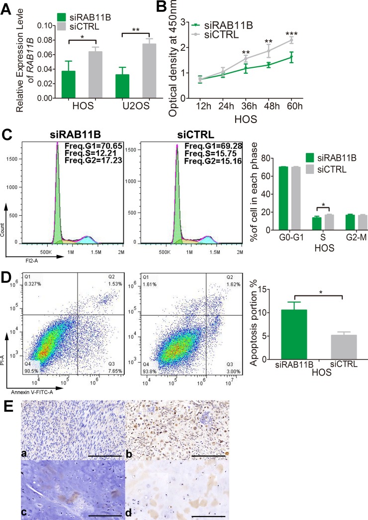
Figure 9. RAB11B promotes osteosarcoma progression.
(A) qRT-PCR analysis of RAB11B in HOS cells and U2OS cells with disrupted RAB11B or not. (B) Proliferation of HOS cells with disrupted RAB11B or not was determined by CCK-8 assay. (C) Flow cytometer analysis of cell cycle distribution in HOS cells with disrupted RAB11B or not. (D) HOS cells with disrupted RAB11B or not were subjected to apoptosis analysis. (E) Analysis of RAB11B expression in osteosarcoma tissues (n = 145) by immunochemistry. Scale bar, 100 μm. RAB11B positive cells were stained brown. (a, b) showed RAB11B expression in bone tissue and (c, d) presented RAB11B expression in cartilage tissue. Data was presented as mean ± SD. The results were reproducible in three independent experiments. *P < 0.05, **P < 0.01, ***P < 0.001.

