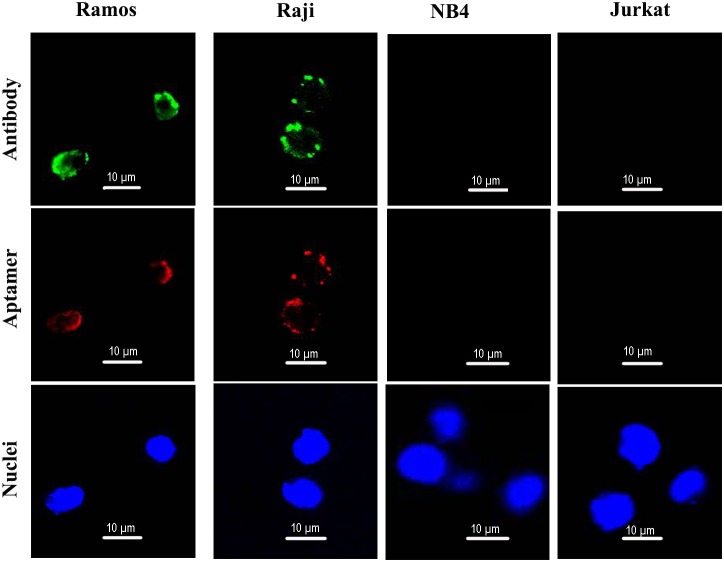
Figure 4. Confocal microscopy evaluation of aptamer's binding to CD19-positive and -negative cells.
Ramos and Raji are CD19-positive cell lines. NB4 and Jurkat are CD19-negative cells. Green fluorescence signal was generated by Alexa Fluor 488-labeled CD19 antibodies. Red fluorescence signal was generated by Cy3-labeled aptamers. The nuclei were stained blue with DAPI.

