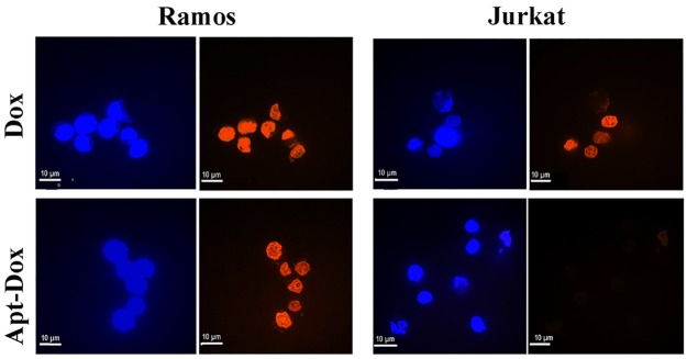
Figure 7. Evaluation of cellular uptake of doxorubicin.
Confocal microscopy images of CD19-positive Ramos cells and CD19-negative Jurkat cells treated with free Dox (upper panel) or Apt-Dox (lower panel). Free doxorubicin emits a red fluorescence. In the lower panel, the red fluorescence was presumably from free doxorubicin released from Apt-Dox, which was taken up by the cells and digested. The nuclei were stained blue with DAPI.

