. 2016 Jan 21;7(5):3017–3024. doi: 10.1039/c6sc00036c

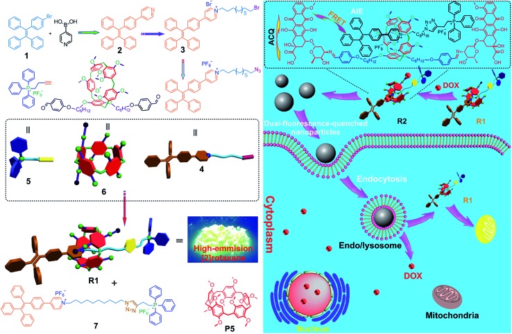
Scheme 1. (Left) Synthetic route to R1 and the corresponding dumbbell-shaped 7 and chemical structures of some compounds used here. (Right) Schematic illustration of the preparation of a mitochondria-targeting probe-inspired prodrug R2 and possible cellular pathways of the dual-fluorescence-quenched R2 nanoparticles. The fluorescences of both TPE and DOX in the nanoparticles self-assembled from R2 are inactivated by the energy transfer relay (ETR) effect, mediated by Förster resonance energy transfer (FRET) and aggregation-caused quenching. The ETR between DOX and R1 is interrupted inside the endo/lysosomes arising from the hydrolysis of the imine bonds. The hydrolyzed DOX and R1 escape from endosomes/lysosomes, move through the cytoplasm toward the nucleus and mitochondria, and cross the mitochondrial membrane and the nuclear membrane, respectively, and the “silenced” fluorescence “wakes up”.

Scheme 1