-
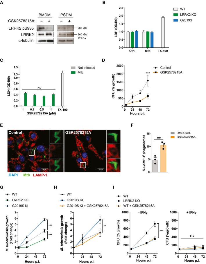
A
Confirmation of efficient LRRK2 kinase inhibition by GSK2578215A 24 h after infection of BMDM or iPSDM with Mtb. Representative image of whole cell lysate Western blotted for LRRK2 pS935, total LRRK2 and α‐tubulin.
-
B, C
LDH assay performed at 72 h post‐infection as toxicity control for CFU experiments in Fig
1. Data show mean ± SD from three technical replicates. LDH assays were routinely performed for CFU and ELISA experiments.
-
D
CFUs in RAW264.7 cells pre‐treated with GSK2578215A (1 μM) or DMSO control for 2 h and infected with Mtb‐eGFP (MOI = 1). *P < 0.5, ***P < 0.001 by Student's t‐test corrected for multiple comparison. Data show mean ± SD. One out of two experiments shown.
-
E
RAW264.7 cells were pre‐treated with 1 μM GSK2578215A or DMSO (Control) for 2 h, and LAMP‐1 recruitment at 24 h post‐infection was assessed using confocal microscopy.
-
F
Quantification of panel (E). Data show mean ± SEM from three independent experiments. **P < 0.01 by Student's t‐test.
-
G
Mtb growth analysed by single cell imaging in WT, LRRK2 KO and LRRK2 G2019S KI BMDM. Data show mean ± SEM from three independent experiments. ***P < 0.001 by Student's t‐test corrected for multiple comparisons.
-
H
Mtb growth analysed by single cell imaging in WT, LRRK2 G2019S KI BMDM and LRRK2 G2019S KI BMDM treated with GSK2578215A for 2 h. Data show mean ± SEM from three independent experiments. **P < 0.001 by Student's t‐test corrected for multiple comparisons.
-
I
CFU in WT BMDM, WT BMDM treated with GSK2578215A and LRRK2 KO BMDM left untreated or pre‐activated with IFN‐γ (100 U/ml over night). Data show mean ± SD from technical replicates. One representative experiment out of three experiments shown. ***P < 0.001 by Student's t‐test corrected for multiple comparison.