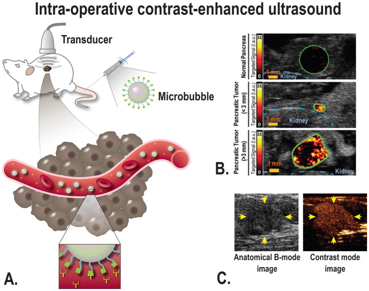
FIGURE 2.
A, Schematic overview of the principle of ultrasound molecular imaging. A molecularly-targeted contrast agent (microbubble) is administered intravenously into the subject (in this case a mouse). Sound waves are transmitted into the subject by the transducer, the sound wave reflections are recorded and converted into images. Because of the size of microbubbles of several micrometers, the contrast agent remains intravascular and attaches to the target of choice (for example VEGFR2). Examples of in vivo molecular ultrasound images with microbubbles in (B) transgenic mouse model of PDAC, showing a strong signal when targeting VEGFR2 in focus of PDAC compared to normal pancreatic tissue, even in small PDAC lesions [From Pysz et al, 201576], and (C) in human with breast cancer using microbubbles targeting kinase insert domain receptor (MBKDR). Left panel: the anatomical image for reference, right panel: MBKDR accumulation in breast cancer lesion [Willmann et al, 201778]

