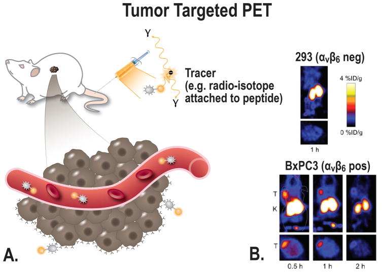
FIGURE 3.
A, Schematic overview of the principle of tumor-targeted PET imaging, a suitable tracer will be administered into the subject (in this case a mouse). Depending on the size of the tracer, the tracer can target the cancer at multiple locations; e.g. intravascular, receptors on the cell membrane, or intracellular. B, Small-animal PET imaging. BxPC-3 (integrin αvβ6 pos) and 293 (integrin αvβ6 negative) cells were xenografted in nude mice. PET images were acquired in tumor-bearing mice using a αvβ6-targeted cysteine knot (18F-fluorobenzoate-R01) [From Hackel et al, 201391].

