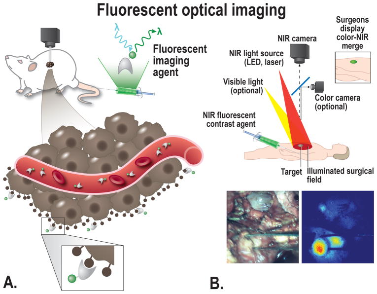
FIGURE 4.
A, Schematic overview of the principle of fluorescent imaging, a suitable targeted imaging agent with a fluorescent dye will be administered into the subject (in this case a mouse). The agent is visualized using a fluorescence imaging system, with an adequate excitation laser and camera able to detect the emitted light. The targeted agents migrate to the cellular targets to visualize the tumor in a target-specific manner, the imaging agent can target the cancer at multiple locations depending on the size; e.g. intravascular, receptors on the cell membrane, or intracellular. B, Top: schematic overview showing the principle of fluorescent imaging. Bottom: Intraoperative image showing the use of tumor-targeted fluorescent guided imaging during pancreatic cancer surgery.

